Back Of Neck Anatomy : Anatomy Of Male Back And Neck Pain In Blue Stock .... The majority of these nerves control the functions of the upper extremities and allow you to feel your arms, shoulder, and back of your head. The cervical spine has 7 stacked bones called vertebrae, labeled c1 through c7. The neck is the start of the spinal column and spinal cord. C7 is the transition with the lumbar vertebrae and has many occipital artery back of neck. Atlas of the anatomy of the head and neck on a ct in axial, coronal, and sagittal sections, and 3d images.
It runs down the back part of the neck, and opens into the external jugular vein just below the middle of its course. Anatomy of the head and neck. Find this pin and more on tips and tricks by wholesome homes. 12 photos of the anatomy of the back of the neck. If you have a back injury, the picture below will help you understand exactly where your problem is.
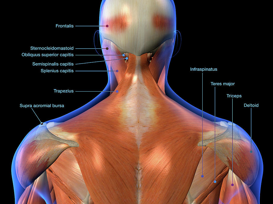
Labeled Anatomy Chart Of Neck And Back Photograph by Hank ... from images.fineartamerica.com This may manifest with both poor head and neck extension, with patients appearing to 'look at the ground.' in these patients, this damage can be a significant cause of. This article describes the anatomy of the head and neck of the human body, including the brain, bones, muscles, blood vessels, nerves, glands, nose, mouth, teeth, tongue, and throat. Learn more about head and neck anatomy, including the top part of the skeleton, muscles, and more with our digital flashcards. This mri neck axial cross sectional anatomy tool is absolutely free to use. Sternocleidomastoid muscle (main muscle in the front of the neck). The longus capitis and rectus capitis anterior are the direct antagonists of the muscles at the back of the neck, serving to restore the head to its natural position after it has been drawn backward. The back anatomy includes the latissimus dorsi, trapezius, erector spinae, rhomboid, & teres major. It comprises the vertebral column (spine) and two compartments of back muscles the anatomy of the head and neck of the human body, including the bones, muscles, blood vessels, nerves, glands, nose, mouth, and throat.
This article concerning the anatomy of the head and neck area gives you a clear structure at hand to see anatomy and function of the cervical organs.
Top head neck anatomy flashcards ranked by quality. The posterior muscles of the neck are primarily concerned with head movements, like extension. The neck is a complex anatomic region between the head and the body. The neck is the part of the body that separates the head from the torso. From the internal carotid, we have two branches, which anastomose with the external carotid via both. So many muscles that cause migraines, arm, neck, shoulders, and back pain. It runs down the back part of the neck, and opens into the external jugular vein just below the middle of its course. This mri neck axial cross sectional anatomy tool is absolutely free to use. During muscle traction, the cheeks are pulled together, which makes food move back and forth between the. Parathyroid glands (glands that control calcium levels in the blood and bones). Find this pin and more on tips and tricks by wholesome homes. The neck is the start of the spinal column and spinal cord. This article covers the anatomy of the deep muscles of the back, including their function, blood supply, innervation, origin and insertion.
Use the mouse scroll wheel to move the images up and down alternatively use the tiny arrows (>>) on both side of the image to move the images. Learn about these muscles, their locations & functional the traps are quite a complex set of muscles. The majority of these nerves control the functions of the upper extremities and allow you to feel your arms, shoulder, and back of your head. Head and upper neck disorders may be called craniovertebral or craniocervical junction anatomy and biomechanics of the craniovertebral junction. The anterior jugular vein (v.
Muscles of the Neck and Torso - Classic Human Anatomy in ... from schoolbag.info They control the scapulae (shoulder blades), which play a role in shrugging, neck movement, head. Call me for an appt. The neck is a complex anatomic region between the head and the body. In the front, the neck extends from the bottom part of the mandible (lower jaw bone) to the bones … in order to fully understand primary neck cancers, it helps to understand the anatomy and function of the structures in the neck. The neck is the area between the skull base and the clavicles. Choose from 500 different sets of flashcards about quiz back anatomy neck muscles on quizlet. The cervical spine has 7 stacked bones called vertebrae, labeled c1 through c7. Jugularis anterior) begins near the.
This article concerning the anatomy of the head and neck area gives you a clear structure at hand to see anatomy and function of the cervical organs.
This mri neck axial cross sectional anatomy tool is absolutely free to use. Parathyroid glands (glands that control calcium levels in the blood and bones). Top head neck anatomy flashcards ranked by quality. Surface anatomy and surface markings bibliographic record list of illustrations subject index. Call me for an appt. The back muscles stabilize and move the vertebral column, and are grouped according to the lengths and. Clinically, surface anatomy is used to split the neck into anterior and posterior triangles which provide clues as to the location of specific structures. The neck is the start of the spinal column and spinal cord. Despite being a relatively small region, it contains a range of important anatomical features. This article concerning the anatomy of the head and neck area gives you a clear structure at hand to see anatomy and function of the cervical organs. The longus capitis and rectus capitis anterior are the direct antagonists of the muscles at the back of the neck, serving to restore the head to its natural position after it has been drawn backward. « back show on map ». Jugularis anterior) begins near the.
So many muscles that cause migraines, arm, neck, shoulders, and back pain. The large spinous process (bump in back of neck) at c7 is called the vertebra prominens. In the front, the neck extends from the bottom part of the mandible (lower jaw bone) to the bones … in order to fully understand primary neck cancers, it helps to understand the anatomy and function of the structures in the neck. Muscle head anatomy vocal organ diagram female neck anatomy neck wireframe head neck human anatomy head artery anatomy face pharynx vector neck degree head anatomy 3d. From the internal carotid, we have two branches, which anastomose with the external carotid via both.
Belayer's Neck - Climbing Magazine from www.climbing.com 12 photos of the anatomy of the back of the neck. The anterior jugular vein (v. The longus capitis and rectus capitis anterior are the direct antagonists of the muscles at the back of the neck, serving to restore the head to its natural position after it has been drawn backward. Sternocleidomastoid muscle (main muscle in the front of the neck). The large spinous process (bump in back of neck) at c7 is called the vertebra prominens. Click now to study the muscles, glands and organs of the neck at kenhub! Choose from 500 different sets of flashcards about quiz back anatomy neck muscles on quizlet. Call me for an appt.
We will attempt to provide a simplified overview of this complex anatomy.
Parathyroid glands (glands that control calcium levels in the blood and bones). Anatomy of the head and neck. Contents of the carotid triangle: From the internal carotid, we have two branches, which anastomose with the external carotid via both. The majority of these nerves control the functions of the upper extremities and allow you to feel your arms, shoulder, and back of your head. C7 is the transition with the lumbar vertebrae and has many occipital artery back of neck. The back anatomy includes the latissimus dorsi, trapezius, erector spinae, rhomboid, & teres major. During muscle traction, the cheeks are pulled together, which makes food move back and forth between the. The neck is connected to the upper back through a series of seven vertebral segments. Want to learn more about it? Learn everything about the neck anatomy with this topic page. In the front, the neck extends from the bottom part of the mandible (lower jaw bone) to the bones … in order to fully understand primary neck cancers, it helps to understand the anatomy and function of the structures in the neck. Find this pin and more on tips and tricks by wholesome homes.
Share :
Post a Comment
for "Back Of Neck Anatomy : Anatomy Of Male Back And Neck Pain In Blue Stock ..."


Post a Comment for "Back Of Neck Anatomy : Anatomy Of Male Back And Neck Pain In Blue Stock ..."